Dedicated
Teleradiology
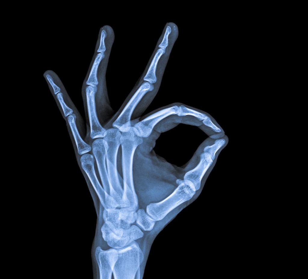
Professional Radiology Outcomes (PRO) is an Australian private company established in 2009. We are a pure teleradiology business focused on Medical and Specialty Radiology Reporting.
We provide offsite radiology reporting services across all modalities and all medical specialties to imaging centres, health departments and hospital groups.
Fast and professional radiology
reporting in 4 simple steps
We invite any Health Professional or Imaging Practice to use our online upload service to upload high resolution patient images easily and quickly.
For routine cases, we will send you a detailed radiology report within 24 hours and can accommodate medical urgent requests even faster.

Upload your images
Complete the form
Make Payment
Receive your report
Experience a
comprehensive
range of services


Our Difference
Our business has no borders, and our service has no limits. Every report represents a human being, and our motivation comes from our desire to deliver the best possible health outcomes.


Hear it from the clients themselves
diagnostic tools to achieve better patient health outcomes.
Here is what some of our clients are saying….
PRO saved me over $1,000 per week in wages by administering my Medicare Compliance & Billing and significantly increased the speed of cashflow into my bank account! Thanks team PRO!
Thanks again for another prompt x-ray report PRO. Can’t recommend your team more.
‘PRO’s service has proved to be a trusted, proactive, and reliable source for our Imaging Practice and our referrers. Nothing we ask of you is refused and makes our jobs that much easier. Thank you!
Our Frequently Asked Questions
What is the difference between PROPACS and ‘one off uploads?
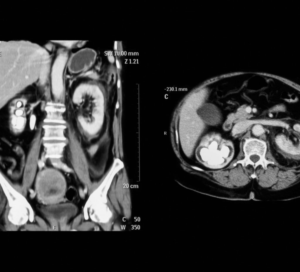
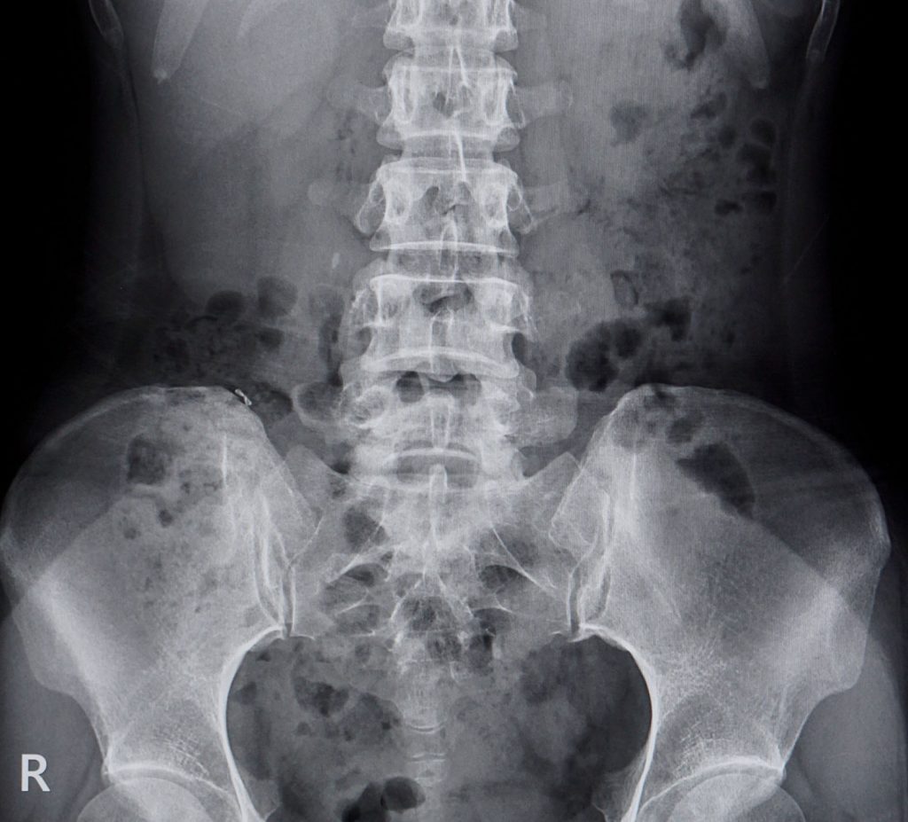
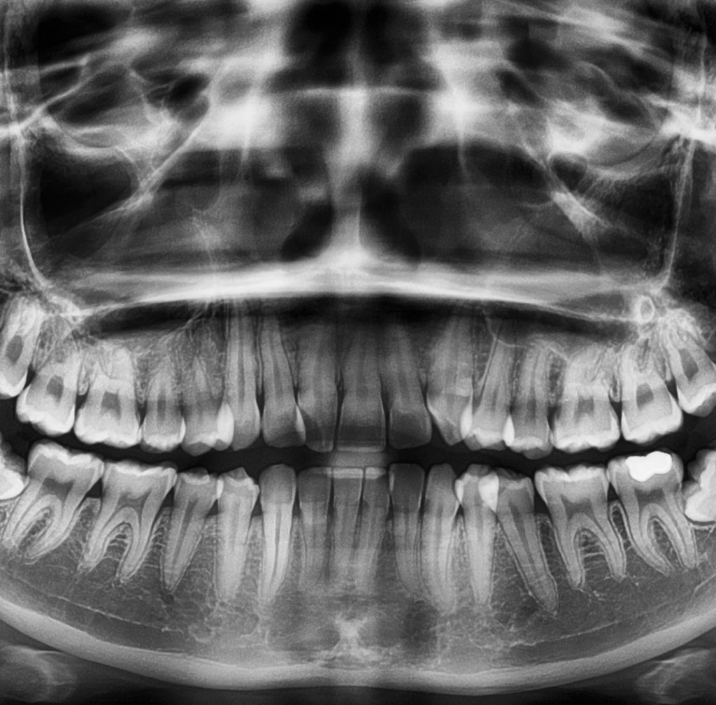
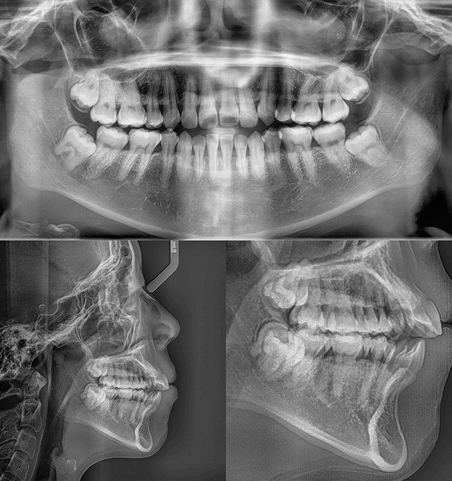
What Modalities do you report?
What is the turnaround time for reporting?
Can referring healthcare providers speak with your radiologists?
As a PROPACS client, will my images be secure?
Preferred Rates for
PROPACS clients
As a PROPACS client, we provide a cost effective teleradiology platform ensuring the best possible patient outcomes 24 hours a day, 365 days per year.